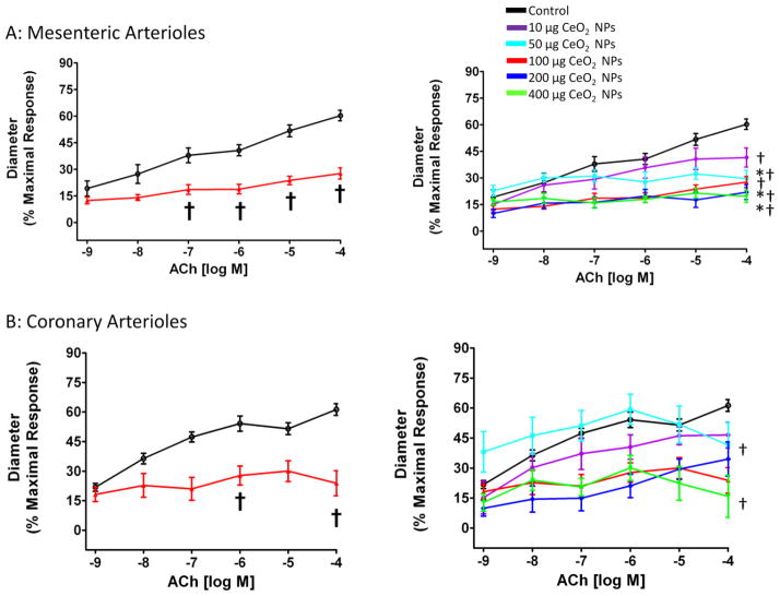
Figure 4.
ACh-induced vasodilation was impaired in mesenteric (A; n=8–13) and coronary (B; n=7–9) arterioles from groups 24 hr post-exposure to CeO2 NPs. Values are means ± SE. † p ≤ 0.05 vs. control; * p ≤ 0.05 vs. 10 μg CeO2 NPs. The right panel represents the responses of the various doses of CeO2 NPs and was analyzed by nonlinear regression. The left panel highlights the point-to-point differences between the control and CeO2 NP exposed group.

