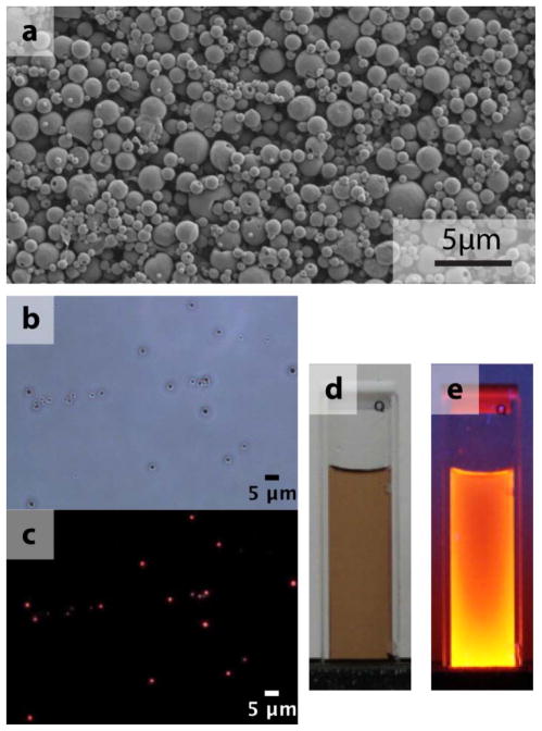
Figure 4.
(a) SEM image of a collection of InvaCuInSexS2-x particles, (b) an optical microscope image in bright field and (c) a corresponding fluorescence image (excitation 480 – 550 nm, emission >590 nm). (d) Photographs show the CuInSexS2-nanocrystals dispersed in toluene in room light and (e) under UV exposure.

