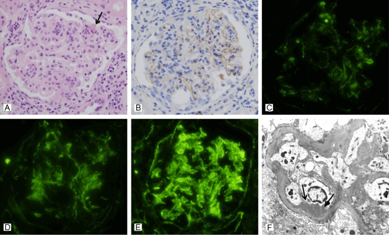
Figure 2.

A representative renal biopsy specimen showing activation of the classical pathway in lupus nephritis (case No. 1). A: Light microscopy in class IV LN shows increased mesangial cellularity with focal lobular accentuation, hyaline thrombi (arrow) and glomerular leukocytic infiltration. A greater degree of periglomerular interstitial inflammation is also noted (hematoxylin and eosin, x 400). B: Moderate glomerular C4d staining in the same case is observed along the glomerular capillary loops and in the mesangium (anti-C4d, x 400). C-E: Immunofluorescence microscopy in the same case reveals granular deposition of C1q (C), C3 (D) and IgG (E) both in the measangium and in the peripheral capillary walls (original magnification x 400). F: Electron microscopy in the same case shows subendothelial deposits along the capillary loops. Loss of foot processes is also observed (transmission electron microscopy, x 8,000).
