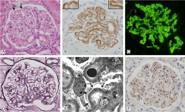
Figure 3.

Glomerular C4d staining in class V membranous lupus nephritis (case No. 4). A: Light microscopy in class V membranous LN shows diffuse thickening of the capillary walls or “wire loop” lesions (arrow) (hematoxylin and eosin, x 400). B: Intense glomerular C4d staining in the same case shows a uniformly strong, diffusely intense and coarsely granular pattern along the glomerular capillary loops, represented by +++. Inset: C4d deposits along the glomerular capillary loops resemble the spikes (anti-C4d, x 400). C: Immunofluorescence microscopy in the same case reveals a granular pattern of IgG deposition in the peripheral capillary walls (anti-IgG immunofluorescence, x 400). D: Markedly thickened glomerular basement membrane and small spikes are seen. Inset: Well-developed spikes are present (periodic acid methanemine silver, x 400). E: Electron microscopy in the same case reveals numerous subepithelial deposits (arrow) scattered throughout the capillary loops (transmission electron microscopy, x 8,000). F: The pattern of glomerular C4d staining in membranous nephropathy (positive control) is similar to the C4d staining pattern in case No. 5 class V membranous LN (anti-C4d, x 400).
