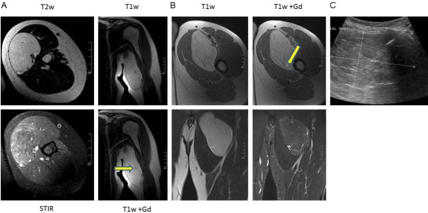
Figure 1.

A: 46 year old woman with hibernoma of the left arm. T1-w and T2-w images show a well circumscribed lesion with a high SI similar to the subcutaneous fat. Within the lesion fine septa can be delineated and a small contrast enhanced vessel (arrow T1-w Gd axial). On STIR images, the hibernoma has higher and more inhomogeneous SI as compared to subcutaneous fat. B: 47 year old man with hibernoma of the left thigh. T1-w and T2-w images show a well circumscribed lesion with a high SI similar to the subcutaneous fat. Within the lesion fine septa can be delineated with a small spotty area of contrast enhancement (arrow T1-w Gd axial). On STIR images, this hibernoma demonstrates similar SI like subcutaneous fat with high signal intensity streaks of fibrovascular tissue. C: The ultrasound image demonstrates a well circumscribed slightly inhomogeneous mass with a biopsy needle in place. The small arrow points to a vessel within a septum.
