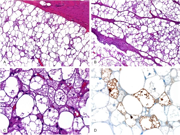
Figure 3.
Histological features of hibernoma. A: Tumor (lower) is well demarcated from adjacent skeletal muscle (upper) by a thin fibrous capsule. Note admixture of pale and granular eosinophilic cells. B: Prominent fibrovascular septa were seen in this case. C: Higher magnification of eosinophilic cells with lipoblast-like appearance. D: Protein S-100 mainly highlighted the microvesicular brown cells.

