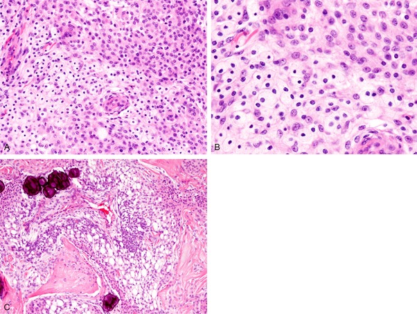
Figure 2.
Histopathological features. A: The tumor comprises two components. The conventional meningioma component (upper right) is composed of polygonal cells with eosinophilic cytoplasm and bland round to oval nuclei. The xanthomatous component (lower left) comprises vacuolated clear cytoplasm with bland round nuclei. HE, x 200. B: Xanthomatous component. The tumor cells have rich clear vacuolated cytoplasm. HE, x 400. C: A transition between the conventional meningioma and the xanthomatous area is present. Psammomas are present. HE, x 100.

