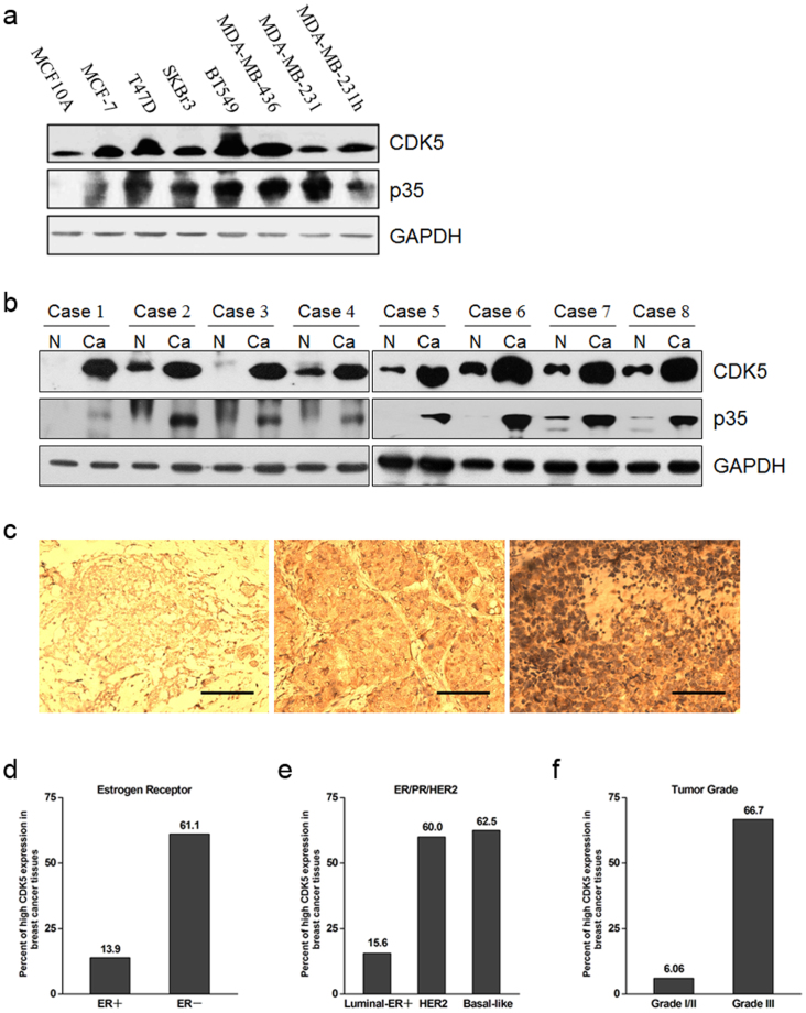
Figure 1. Enhanced expression of CDK5 and p35 in breast cancer cells and cancerous tissues.
(a) immunoblots of CDK5 and p35 protein expression in MCF10A mammary epithelial cells and breast cancer cells. (b) immunoblotting analysis of CDK5 and p35 protein expression in breast cancer tissue specimens, including non-cancerous surrounding tissues (N) and cancerous tissues (Ca). (c) immunohistochemistry of CDK5 protein in breast cancer tissue specimens. Left, weak staining; middle, moderate staining; right, strong staining. Scale bar = 100 μm. (d), (e) and (f) percentages of human breast cancer specimens with high level of CDK5 expression in different tumor subtypes and different tumor grades. Corresponding p-values analyzed by χ2 test are indicated.

