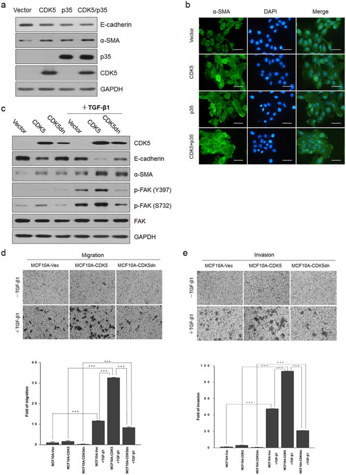
Figure 4. CDK5 had a potential synergistic effect on TGF-β1-induced EMT via CDK5 kinase activity on the phosphorylation of FAK at Ser-732.
(a) immunoblotting analysis of expression of CDK5 and p35, the epithelial marker E-cadherin, and the mesenchymal markers N-cadherin and α-SMA in MCF10A-Vector, MCF10A-CDK5, MCF10A-p35 and MCF10A-CDK5-p35 cells. (b) immunofluorescence staining for the mesenchymal marker α-SMA. Scale bar = 50 μm. (c) immunoblotting analysis of expression of CDK5, the epithelial marker E-cadherin, the mesenchymal markers α-SMA, FAK, p-FAK Y397 and p-FAK S732 in MCF10A-Vector, MCF10A-CDK5 and MCF10A-CDK5dn cells with or without TGF-β1 treatment. (d) and (e) migration (24 h; d) and invasion (60 h; e) assays in MCF10A-Vector, MCF10A-CDK5 and MCF10A-CDK5dn cells with or without TGF-β1 treatment. The mean was derived from cell counts of 5 fields, and each experiment was repeated 3 times (***, P < 0.001, compared with the control). Representative images of migrated and invaded cells are shown (upper).

