
Figure 8. Cav3.2 immunostaining was found within the dermal-epidermal layer of glaborous skin from the mouse hind paw.
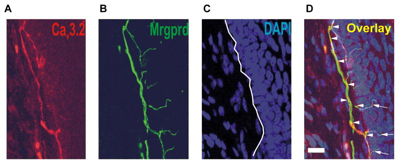
Confocal microscopy revealed that expression of CaV3.2-positive fibre (red) was found within the dermal-epidermal border of Mrgprd-KI mouse hind paw (E). Panel F shows Mrgprd positive fibres (green) in the same slide as panel A. Panel C of this figure shows outlines of cell’s nuclei on same slide stained with DAPI (blue). Panel H shows overlay of staining (yellow) depicted on panels E, F and G. Note that Cav3.2 immunostaining was largely co-localized with Mrgprd staining. Scale bars = 20 μm.