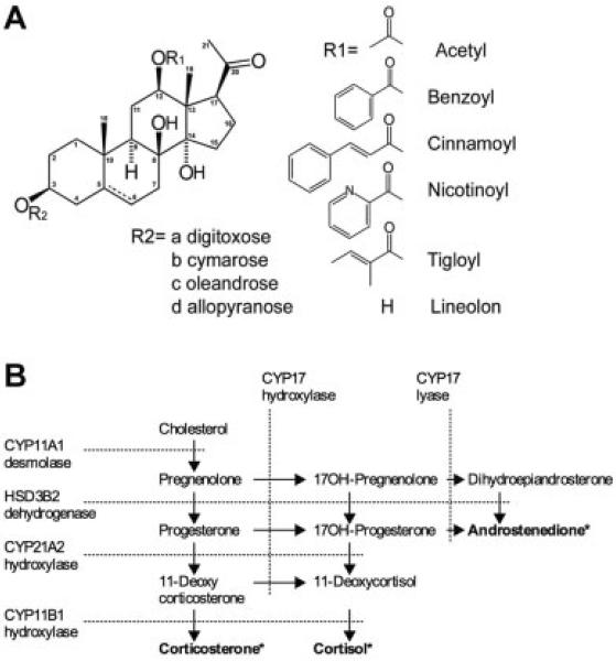
Fig. 1.
Pregnane glycosides and steroidogenesis. A: Chemical structure of pregnane glycosides used in this study showing a common steroid core, C12 side chain moieties, deoxysugar moieties forming polysaccharide glycosylation pattern at the C3 carbon, and an optional double bond between C5 and C6 carbons. B: Schematic illustration of steroidogenesis in human adrenal H295R cells showing end-point steroidogenic metabolites (*), metabolite conversion pathways (arrows), and respective enzyme that catalyze them (dashed lines).

