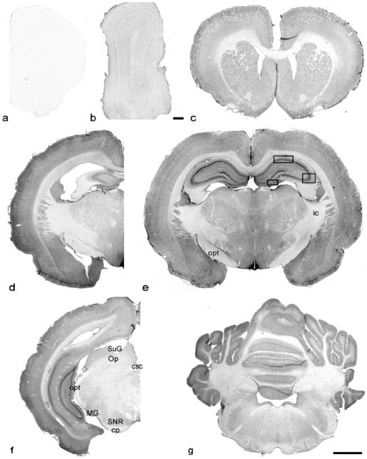
Fig. 1.
Immunolabeling of NR1 in coronal sections of AGS brain from forebrain to cerebellum: (a) control section at the same level as the NR1 stained section shown in (c); (b–f) olfactory bulb to cerebellum; (a–c), (d) and (f) are from ibeAGS; (e) and (g) are from hAGS. opt, optic tract; ic, internal capsule; csc, commissural of the superior colliculus; SuG, superficial gray layer of the superior colliculus; Op, optic nerve layer of the superior colliculus; MG, medial geniculate nucleus; SNR, substantia nigra, reticular part; cp, cerebral peduncle, basal part. Scale bar in (b), 100 μm, in others, 2.5 mm. Frames in (e) indicate the areas where neuronal soma size was measured.

