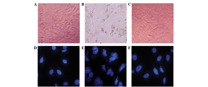
Figure 1.
Morphological post-irradiation changes of the HEI-OC1 cells. (A) HEI-OC1 sham-irradiated cells showing a polygon and fusiform morphology. (B) The HEI-OC1 cells following 16-Gy irradiation for 72 h showing a dendritic or amoeboid appearance, with numerous highly ramified processes and body swelling. (C) The majority of the cochlear hair cells treated with tanshinone IIA and irradiation show a polygon and fusiform morphology, rather than a dendritic or amoeboid appearance. (D) The nuclei of the cells showing a uniform, oval shape following irradiation. (E) The cells were treated with 16 Gy. The cell nuclei exhibit shrinkage, fragmentation, megakaryocytes and deformation. (F) The cells were co-treated with tanshinone IIA and radiation and the nuclei appear to be oval-shaped and of a relatively homogeneous size. Magnification, (A–C) ×200; (D–F) ×400 (4′,6-diamidino-2-phenylindole staining).

