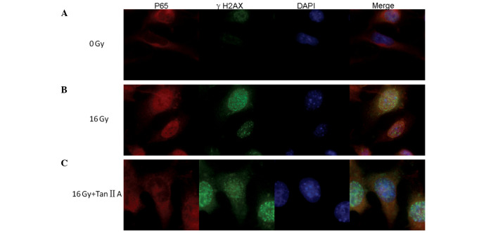
Figure 5.
Post-irradiation expression of γ-H2AX and p65. Confocal images of immunofluorescence double staining for γ-H2AX and p65 in the HEI-OC1 cells following 16 Gy irradiation at 24 h with or without pretreatment with 8 mg/ml tanshinone IIA. DNA was counterstained with DAPI. Original magnification, ×400. (A) HEI-OC1 cell irradiation showing the minimal expression of γ-H2AX and the expression of p65 in the cytoplasm. (B) HEI-OC1 cells following 16 Gy irradiation showing the expression of γ-H2AX foci formation and showing that p65 is translocated to the nucleus. (C) HEI-OC1 cells following 16 Gy irradiation pre-treatment with tanshinone IIA showing the expression of γ-H2AX foci formation and showing that p65 is inhibited from translocating into the nucleus. DAPI, 4′,6-diamidino-2-phenylindole.

