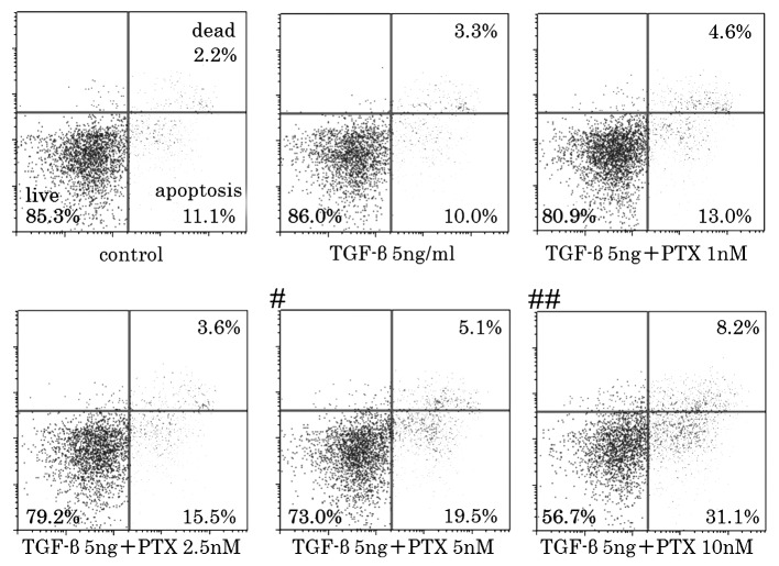
Figure 2.
Flow cytometry of CCKS-1 cell death assay using various concentrations of PTX and/or TGF-β. The percentage of apoptotic/dead cells increased at concentrations of ≥5nM and were significantly increased at ≥10nM. Statistically significant differences were determined by the χ2 test. #P<0.05; ##P<0.01 vs. control. TGF-β1, transforming growth factor-β1; PTX, paclitaxel.

