
Figure 1.
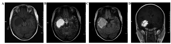
Pre-operative MRI of the initial surgery. (A) A hypointense well-defined lesion in the right temporal lobe, shown by T1WI. (B) A homogeneous hyperintense lesio, shown by T2WI. (C) A heterogeneous hyperintense lesion with slight edema, shown by FLAIR sequence imaging. (D) Patchy enhancement of the tumor, shown by contrast-enhanced imaging. MRI, magnetic resonance imaging; WI, weighted imaging; FLAIR, fluid-attenuated inversion recovery.