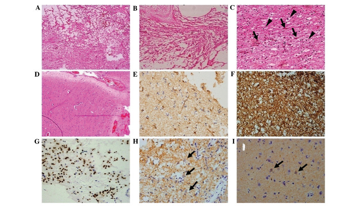
Figure 3.
Pathology data from the first surgery. (A and B) A typical loose reticular degeneration with microcapsule formation and matrix mucoid degeneration (HE staining; magnification, ×4). (C) Hyperplastic oligodendrocyte-like cells (black triangles) and immature neurons (black arrows) in the tumor section (HE staining; magnification, ×40). (D) Evidence of FCD in the peritumoral cortex (HE staining; magnification, ×20). (E) Astrocytoma, (GFAP staining; magnification, ×40). (F) Hyperplastic gliacyte component, including oligodendrocyte-like cells and astrocytoma (S-100 staining; magnification, ×40). (G) Oligodendrocyte-like cells, (Oligo-2 staining; magnification, ×40). (H) Immature neurons (black arrows; Syn staining; magnification, ×40). (I) Immature neurons (black arrows; NF staining; magnification, ×40). HE, hematoxylin and eosin; FCD, focal cortical dysplasia; GFAP, glial fibrillary acidic protein; Syn, synaptophysin; NF, neurofilament.

