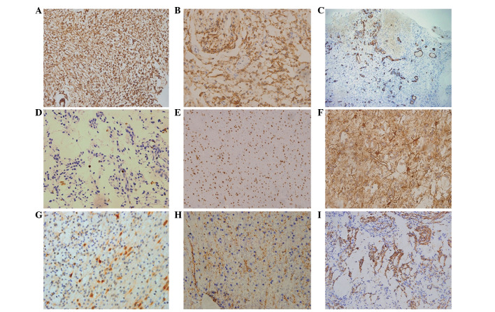
Figure 7.
Pathology data following the second surgery. (A) Hyperplastic gliacyte component (GFAP staining; magnification, ×20). (B) Hyperplastic gliacyte component (S-100 staining; magnification, ×40). (C) Hyperplasia of the capillaries (CD34 staining; magnification, ×4). (D) Rare mitotic figures of the tumor (Ki-67 staining; magnification, ×40). (E) Oligodendrocyte-like cells in the region of the microcapsules (Oligo-2 staining; magnification, ×20). (F) Astrocytoma in the region of microcapsules (GFAP staining; magnification, ×40). (G) Immature neurons (NeuN staining; magnification, ×40). (H) Immature neurons (Syn staining; magnification, ×40). (I) The focal positive reaction of the tumor (NF staining; magnification, ×20). GFAP, glial fibrillary acidic protein; Syn, synaptophysin; NF, neurofilament.

