Figure 1.

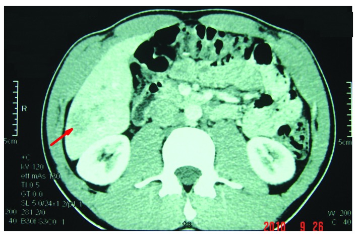
Computed tomography (CT) scan showing the Neoplasm (~3.5×4.5×4.5 cm) in the right lobe of the liver, as indicated by the arrow.

Computed tomography (CT) scan showing the Neoplasm (~3.5×4.5×4.5 cm) in the right lobe of the liver, as indicated by the arrow.