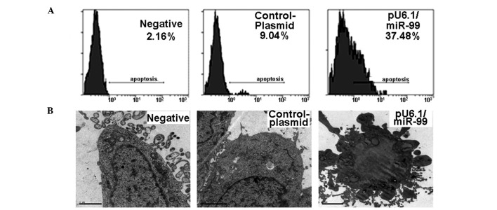
Figure 3.
Apoptotic cells were detected by FACS and electron microscopy. (A) More apoptotic HeLa cells (~37.48%) were observed among pU6.1/miR-99 transfected cultures compared with that among the controls (2.16 or 9.04%). (B) Variations in cell morphology were observed by electron microscopy. Compared with the controls, pU6.1/miR-99-transfected cells showed increases in intracellular electron density and the proportion of nuclear plasma. The cell nucleus presented material densification or plaques.

