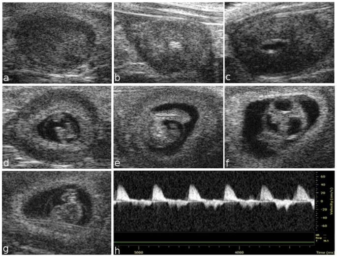
Figure 1. Ultrasound images of fetal development from embryonic day 5.5 to 9.5.
Representative images of the implantation site at E5.5 showing the apposition of the blastocyst trophectoderm with the uterus (a). At E6.5, a small echolucent cavity containing the embryo is visualized inside the decidua (b). At E7.5, the amniotic and exocoelomic cavities and the ectoplacental cone region are discernable (c). At embryonic day 8.5, the heart, head and whole embryo are visualized (d). At E9.5, the amniotic membrane, yolk sac and cerebral ventricles are visible, while a Doppler spectral trace of ventricular inflow and outflow can be observed in the “U shaped” heart tube (e–h).

