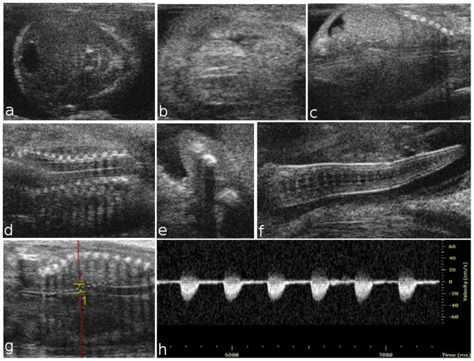
Figure 3. Ultrasound images of fetal development from embryonic day 14.5 to 16.5.
At E14.5, progressive ossification is observed in the skull (a) and ribs (c), and the interventricular septum is completed (b). At 15.5, the development of the vertebral elements and humerus is complete (d, e). At E16.5, the curl is clearly visible (f). Dorsal aorta and corresponding Doppler spectral trace at E16.5, which exhibits a rapid upstroke and return to zero velocity during diastole (g, h).

