
Figure 4. Masson stain of PQ group (×200).
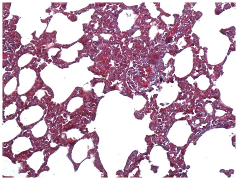
The alveolar wall was significantly thickened, there was evidence of phoroblast hyperplasia and fibroglia fibrils, which were obviously hyperplastic.
Official websites use .gov
A
.gov website belongs to an official
government organization in the United States.
Secure .gov websites use HTTPS
A lock (
) or https:// means you've safely
connected to the .gov website. Share sensitive
information only on official, secure websites.
The alveolar wall was significantly thickened, there was evidence of phoroblast hyperplasia and fibroglia fibrils, which were obviously hyperplastic.