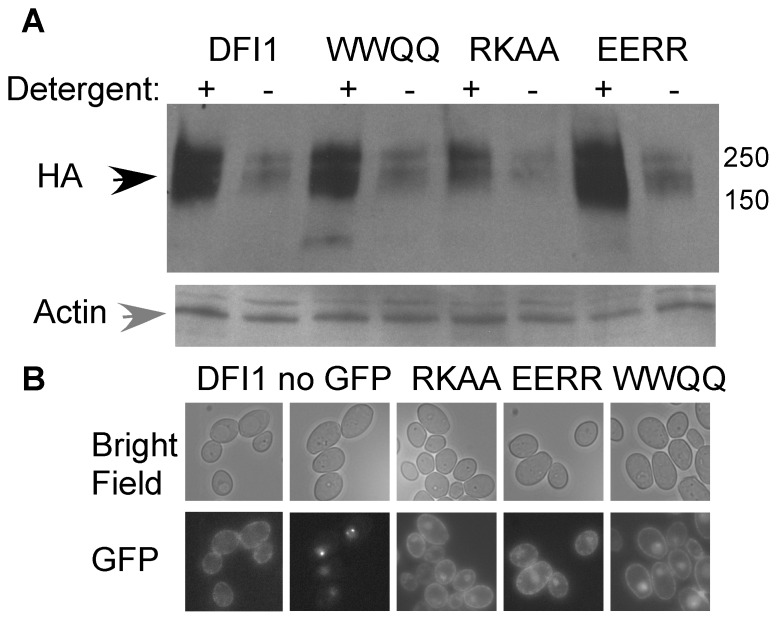
Figure 3. Expression and localization of Dfi1p mutant proteins.
A, Wild type and mutant Dfi1-TAP protein are extractable with detergent. Cells were grown in YPD overnight, collected and total protein extracted in extraction buffer with (+) or without (−) 1% triton-X100 and 0.5% sodium deoxycholate. Equal amounts of total protein were fractionated on an SDS-PAGE gel and Western blotted with anti-HA (top) to detect Dfi1p (black arrow) or anti-actin (gray arrow) as a loading control (bottom). DFI1, DFI1-TAP; WWQQ, dfi1-WWQQ-TAP; RKAA, dfi1-RKAA-TAP; EERR, dfi1-EERR-TAP. Molecular weight markers are shown to the right (in kDa). B, Dfi1-GFP strains show GFP fluorescence at the periphery of the cell. Top, bright field; Bottom, GFP fluorescence. DFI1, DFI1-GFP; no GFP, Δdfi1 null with no GFP; RKAA, dfi1-RKAA-GFP; EERR, dfi1-EERR-GFP; WWQQ, dfi1-WWQQ-GFP.

