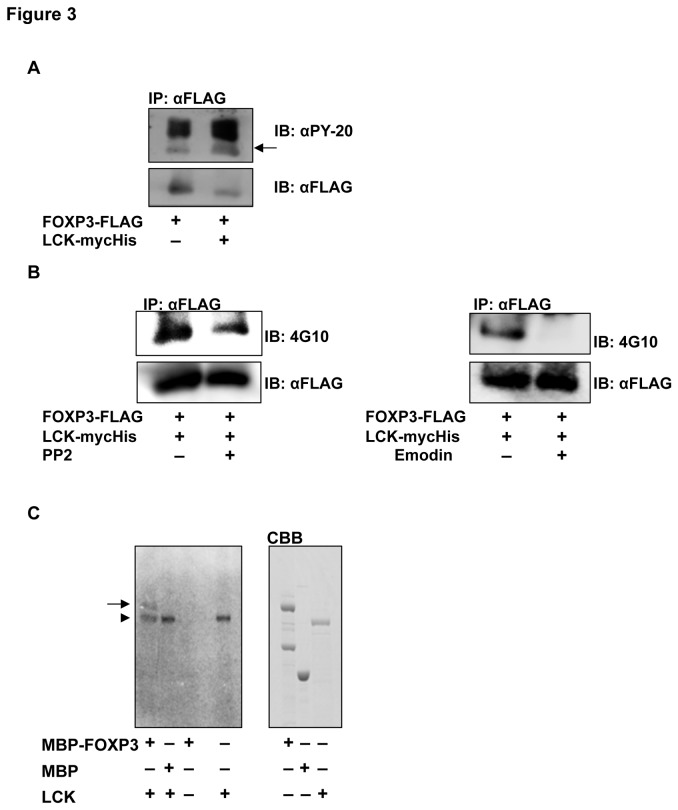
Figure 3. FOXP3 phosphorylation by LCK.
(A) Phosphorylation of FOXP3 in MCF-7 cells. FOXP3 was immunoprecipitated with an anti-FLAG antibody and immunoblotted with an anti-PY-20 antibody (top). The antibodies were stripped and FOXP3-FLAG was detected using an anti-FLAG antibody (bottom). FOXP3 co-expressed with LCK was potently phosphorylated (arrow) compared with only FOXP3. (B) Decreased phosphorylation of FOXP3 by LCK inhibitors, PP2 (left) and emodin (right). Phosphorylated (top) and total (bottom) immunoprecipitated FOXP3 were detected using the indicated antibodies. Both PP2 and emodin inhibited the phosphorylation of FOXP3. (C) In vitro kinase assay. The recombinant proteins were incubated and separated using SDS-PAGE, and then autoradiographed (left). Right panel indicates each recombinant protein stained with CBB. Phosphorylated MBP-FOXP3 (arrow) was detected in the lane containing MBP-FOXP3 and LCK (an arrowhead).

