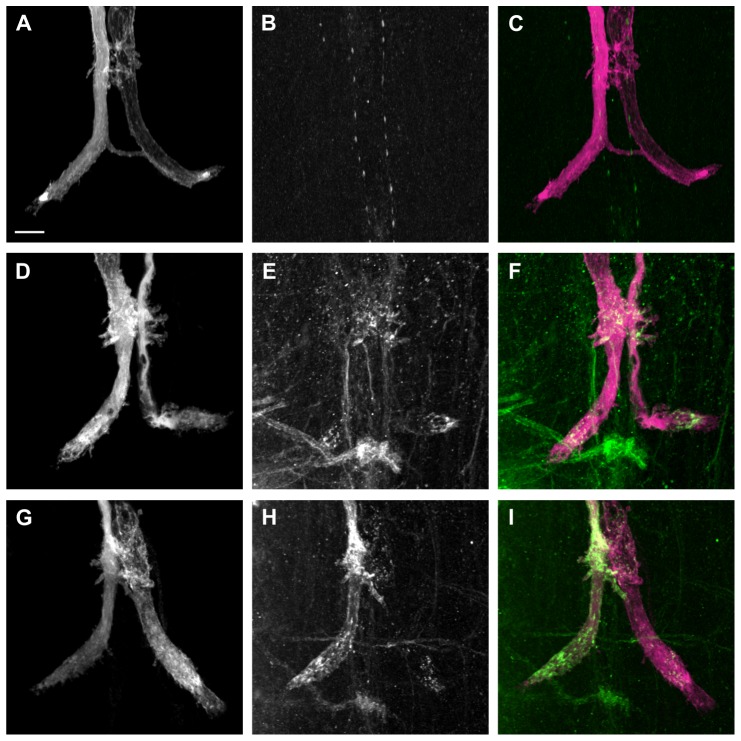
Figure 5. Immunohistochemical labeling of L1-C264Y at GF terminals.
Alexa Fluor 555 dye injections were performed to label the GFs (magenta) and anti-L1 antibody staining is shown in green. Grayscale is used for single staining while the composite images are in corresponding colors. All images are maximum intensity projections from confocal image stacks. Scale bar is 10 µm. (A-C) OK307/+ negative control animal. The fluorescently labeled GFs (A and C, grayscale and magenta) and the ventral nerve cord (VNC) labeled with anti-L1CAM antibody (B and C, grayscale and green). Composite image is shown (C). (D–F) OK307,UAS-L1-C264Y/+ animal. The GFs were labeled with Alexa Fluor 555 (D and F, grayscale and magenta) and the VNC with anti-L1CAM (E and F, grayscale and green). Co-localization of both labels is shown in (F). L1-C264Y protein was detected in both GF terminals at the terminal surface as well as in vesicular clusters in the cytoplasm. (G–I) OK307,UAS-L1-C264Y/+ animal. The overlap of the GFs (G and I, grayscale and magenta) and anti-L1CAM staining in VNC (H and I, grayscale and green) are shown as a composite in (I). In the left GF terminal, L1-C264Y protein was strongly detected at the terminal surface as well as in the cytoplasm. In the right GF terminal, L1-C264Y protein was only labeled weakly in few vesicular clusters inside the axon terminal.

