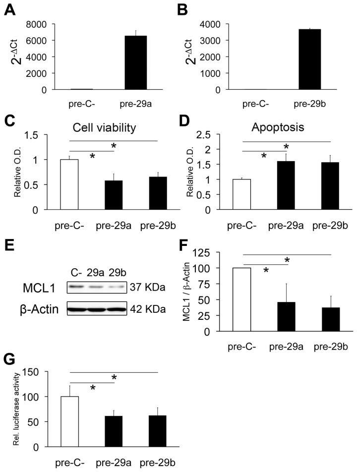
Figure 6. Over-expression of miR-29a/29b targets MCL1 and promotes apoptosis in GICs.
miR-29a/b over-expression in GN1C cells transfected with pre-miR-29a (pre-29a) or pre-miR-29b (pre-29b) compared to pre-miR negative control (pre-C-) was confirmed by q-RT-PCR 4 days after transfection (A, B). Cell viability assays using MTS (C) and apoptosis assessment by Cell Death Detection kit (D) were carried out 4 days after transfection. MCL1 protein levels were also measured by Western blot at the same time point, using β-Actin as loading control (E). Quantification of Western blots was performed with ImageJ (F) and is displayed as the MCL1/β-actin ratio relative to the negative control (100%). Regulation of the 3´-UTR of MCL1 by miR-29a and miR-29b was analyzed by luciferase assays (G). All experiments were carried out at least in triplicate. * indicates p value <0.05 in unpaired t test or Mann-Whitney U test, using the Holm-Bonferroni correction for multiple comparisons.

