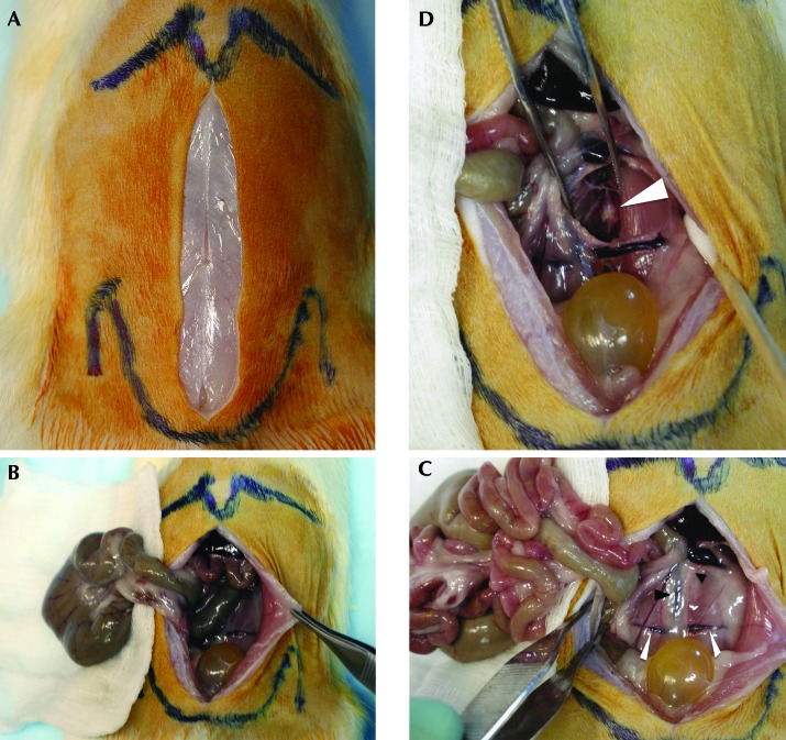
Figure 1.
Photographs from a necropsy dissection simulating surgical technique. (A) The skin incision has been created, and palpated bony landmarks are outlined: xiphoid process projecting distally at the top of the image and iliac wings prominent laterally in vicinity of the pelvis meeting in the midline distally at the pubis. (B) The cecum (large intestine) has been extraperitoneally reflected onto saline-moistened 4×4 gauze pads. (C) The cecum and small intestine have both been reflected extraperitoneally; note the increased visualization of the retroperitoneum and traversing iliolumbar veins (white arrowheads). Black arrowheads indicate the left ureter (small) and aorta–inferior vena cava (large). (D) The ureter and great vessels have been dissected medially and the left psoas laterally; the forceps maintain the deep exposure in the dissection plane, and the arrowhead indicates the ventral disc annulus.

