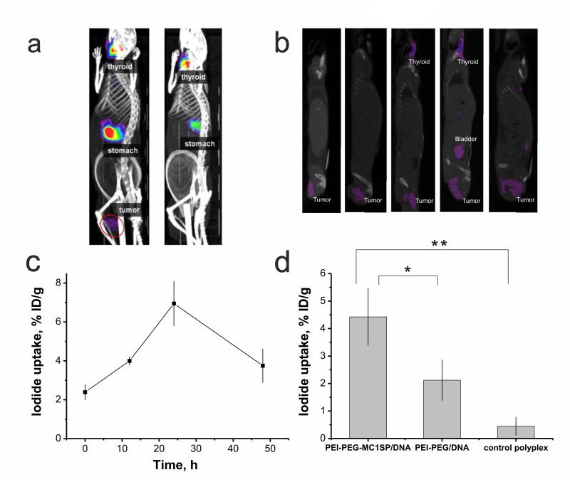
Fig. 6.
SPECT/CT imaging of 123I accumulation in М3 melanoma tumor after NIS gene transfer. Reconstructed 3D-projections of mice at 24 h after injection of targeted polyplexes with plasmid DNA encoding NIS (left) or targeted polyplexes containing plasmid without promoter as a control (right) (a). Reconstructed images of saggital planes passing through the tumors of five mice at 24 h after injection of targeted polyplexes (b). NIS expression with time measured by iodide uptake after intravenous administration of targeted polyplexes, containing 80 μg of plasmid DNA encoding NIS (c). 123I accumulation in tumor at 24 h after injection of targeted (PEI-PEG-MC1SP/DNA) polyplexes compared to non-targeted (PEI-PEG/DNA) ones and targeted polyplexes containing plasmid without promoter (control polyplexes) (d). Background level observed in non-transfected control (2.4 ± 0.4 % ID/g) was subtracted in (d). 18.5 МBq of 123I was injected intravenously for visualizing of NIS gene expression. All values are means ± SD % ID/g. *p < 0.001, **p < 0.0001.

