Figure 1.
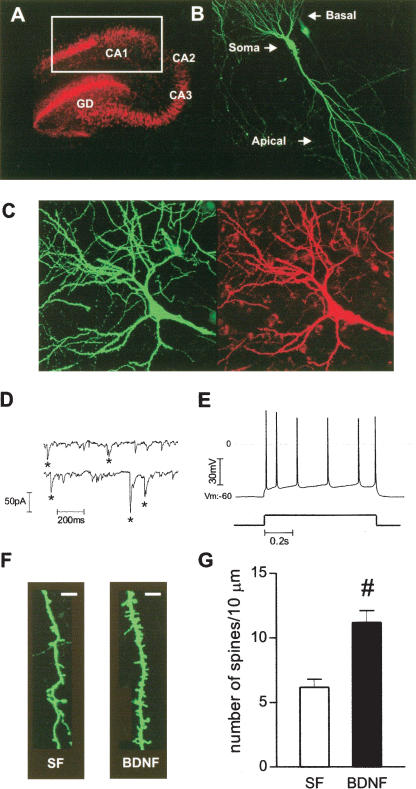
BDNF increases spine density in eYFP-transfected CA1 pyramidal neurons. (A) NeuN immunocytochemistry allowed identification of all hippocampal subfields, CA1, CA3, and dentate gyrus (DG). White square indicates the region from which pyramidal neurons were selected for confocal imaging. (B) Confocal image of a representative eYFP-transfected CA1 pyramidal neuron from a serum-free slice. (C) Confocal image of a representative eYFP-transfected CA1 neuron (green), filled with Alexa-594 during whole-cell recording (red). (D) Representative continuous records of spontaneous excitatory synaptic currents. (E) Action potential train (top trace) evoked by somatic current injection (bottom trace, 200 pA, 0.8 sec). (F) Higher magnification views of representative segments of apical dendrites from control serum-free (SF) and BDNF-treated slices (scale bar, 2 μm). (G) Quantification of spine density, expressed per 10 μm of apical dendrite. (#) P < 0.001, Student t test, t = 4.615, n = 10-13.

