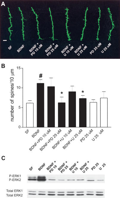
Figure 2.
ERK activity is necessary for BDNF to increase spine density in CA1 pyramidal neurons. (A) High magnification views of representative segments of apical dendrites (scale bar, 2 μm). Images (left to right) are as follows: serum-free (SF); BDNF; BDNF + PD98059 at 10 μM (BDNF + PD 10 μM); BDNF + PD98059 at 25 μM (BDNF + PD 25 μM); BDNF + U0126 at 10 μM (BDNF + U 10 μM); BDNF + U0126 at 25 μM (BDNF + U 25 μM); PD98059 at 25 μM alone (PD 25 μM); and U0126 at 25 μM alone (U 25 μM). (B) Quantification of spine density, expressed per 10 μm of apical dendrite. (#) P < 0.01 vs. SF group; (*) P < 0.05 vs. BDNF-treated group; ANOVA followed by Newman Keul test, F(7, 64) = 3.475; n = 5-13. (C) Representative Western blots using antiphosphorylated ERK1 and ERK2 (P-ERK1 and P-ERK2) and anti-ERK1 and ERK2 (Total ERK1 and Total ERK2) antibodies in whole homogenates of hippocampal slice cultures. Lanes (left to right) are as follows: serum-free (SF); BDNF; BDNF + PD98059 at 10 μM (BDNF + PD 10 μM); BDNF + PD98059 at 25 μM (BDNF + PD 25 μM); BDNF + U0126 at 10 μM (BDNF + U 10 μM); BDNF + U0126 at 25 μM (BDNF + U 25 μM); PD98059 at 25 μM alone (PD 25 μM); and U0126 at 25 μM alone (U 25 μM).

