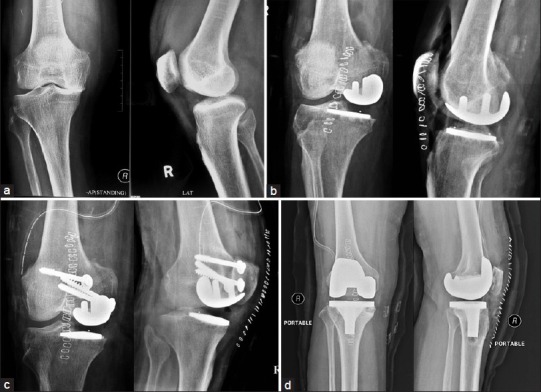
Figure 2.

(a) Preoperative X-ray anteroposterior and lateral views of (R) knee joint (case E02) showing tricompartmental OA of the right knee. (b) Immediate postoperative X-ray anteroposterior and lateral views after UKA surgery showing implant in situ (c) X-ray anteroposterior and lateral views showing screw fixation of medial femoral condyle fracture under the UKA. (d) X-ray after revision to TKA
