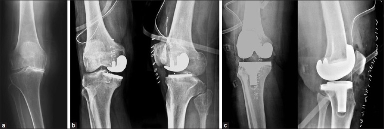
Figure 3.

(a) Preoperative X-ray anteroposterior view (standing) (case E03) showing severe osteoarthritic changes (b) Postoperative X-ray anteroposterior and lateral views following UKA showing possible valgus placement. (c) Postoperative X-ray anteroposterior and lateral views after revision to TKA
