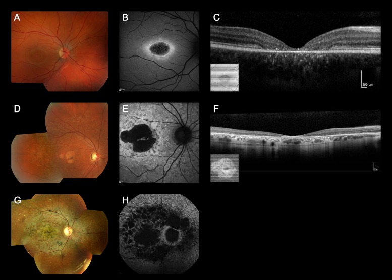
Figure 1.
Color fundus photographs, autofluorescence images, and optical coherence tomography of three representative cases harboring two or more ABCA4 variants with “typical” ABCA4-associated retinal disease (patients 19, 36, and 17). Color fundus photograph of patient 19 shows macular atrophy (A) and AF imaging demonstrates a localized low AF signal at the fovea, with a high signal edge surrounded by a homogeneous background (B). SD-OCT demonstrates marked outer retinal loss at the central macula (C). Patient 36 has macular atrophy surrounded by numerous yellow-white flecks (D) and a localized low AF signal at the macula surrounded by a heterogeneous background, with peripapillary sparing (E). Generalized loss of outer retinal architecture is seen on SD-OCT. Patient 17 has widespread multiple areas of atrophy with patchy pigmentation (G) and multiple areas of low AF signal at the posterior pole with a heterogeneous background (H).

