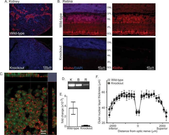
Figure 1.
Klotho is expressed in retina. (A) Representative images of kl protein expression detected by AF1819 in wild-type but not knockout mouse kidney. Nuclei (blue) detected by DAPI. (B) Representative images of kl protein expression (red) by detected by AF1819 in wild-type but not knockout mouse retina. Nuclei detected by DAPI. (C) Confocal microscopy with orthogonal projections to detect klotho protein expression (red) and DAPI (represented as pseudo color green, nucleus) in a single optical section (0.35 μm) from the INL of a wild-type retina. (D) Endpoint PCR product detected on ethidium bromide stained gels indicating relative expression levels of kl mRNA in wild-type mouse kidney (K), brain (B), and retina (R). (E) The fold change of kl mRNA expression in wild-type and knockout mouse retina. Messenger RNA was isolated from retina and kidney and expression level measured by Taqman qPCR. Fold change calculated as 2−ΔΔCt as discussed in methods. (F) Morphometric analysis at 7 weeks of age revealed no evidence of degeneration in kl knockout retina as outer nuclear layer thickness was indistinguishable from wild-type retina. (n = 6 per group, ±SEM). OS, outer segment; IS, inner segment.

