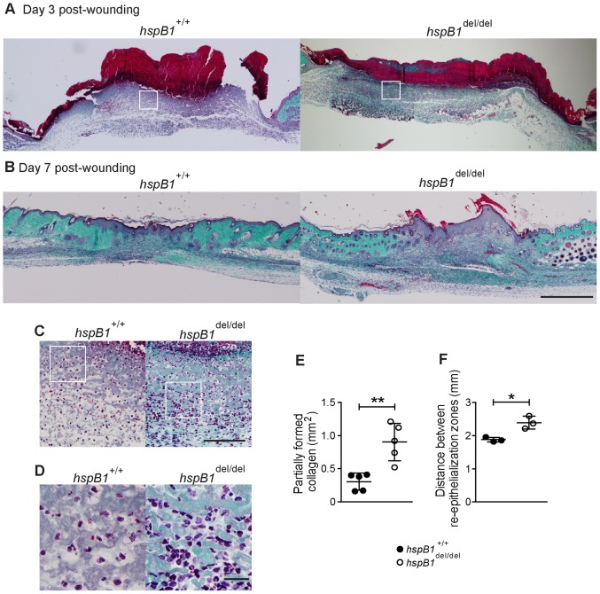
Figure 6. Histological analysis showing reduced re-epithelialisation, impaired collagen deposition and increased cellular infiltration in hspB1 del/del relative to wild-type wounds.
A, Masson’s trichrome staining of wild-type and hspB1 del/del d3 and B, d7 wounds as in Fig. 5; bar = 500 µm. C, High power images of cellular infiltrate from boxed regions indicated in (A) are shown (bar = 100 µm). D, as for (C) but at higher magnification showing cells with multi-lobed nuclei characteristic of neutrophils; (bar = 20 µm).E, Plot showing mean areas of incomplete collagen deposition in d7 wild-type and hspB1 del/del wounds (n = 5); **P<0.01. F, Plot of mean distance±SEM (n = 3 mice per group) between re-epithelialisation margins (re-ep) in wild-type and hspB1 del/del mice in d3 wounds; (*P<0.05 calculated by Student’s t-test of 12 individual wounds per group from 3 experiments). 11–14 week age-matched female wild-type and hspB1 del/del mice were used.

