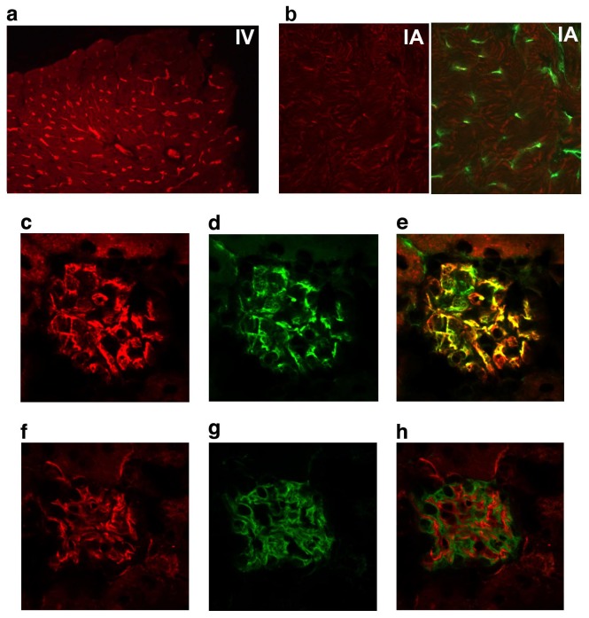
Figure 1. MOA lectin binds the glomerular endothelium.
Intravenous injection of MOA lectin labels heart microvascular endothelium (A), but after intra-arterial injection, the kidney glomerular (C, F) but not heart endothelium (B) is labeled. Endothelial cells are labeled with anti-CD31 (green; B (right panel), D), or MOA lectin (red; A, B (left panel), C, F). Double-labeled merged images of CD31 and MOA lectin (E) demonstrate overlapping distribution in the glomerulus. No overlap is identified between MOA lectin (F) and the podocyte marker, podocin (G) in the merged image (H).

