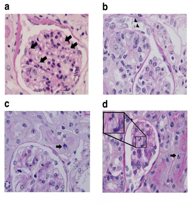
Figure 4. Glomerular microvascular injury after sublethal LPS/ LS treatment.

Mice were treated with LPS/ LS 200 μg/kg and tissues were harvested 4 days later and stained with PAS. Apoptotic cells (arrows) are seen in the glomerular capillaries (A) and tubules (B) after LS treatment. Regenerative mitotic changes are evident in the tubular (C) and glomerular capillary compartments (D).
