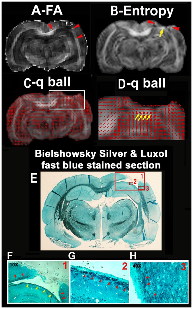
Figure 7. The FA (A), diffusion entropy (B), q-ball fiber orientation (C, D) maps overlayed onto entropy, and the Bielshowsky and Luxol fast blue immunoreactive staining images (E-H) measured from the fixed animal brain.

The images in F, G are high magnification images from the box areas in image E as indicated by the numbers in the up right corner in the images of E and F.
