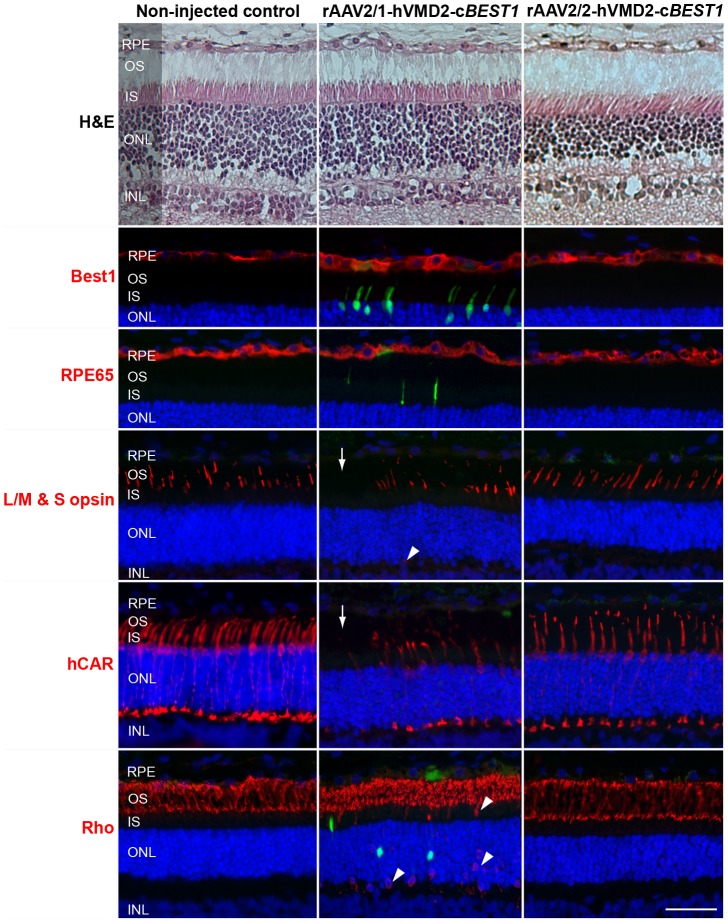
Figure 6. Consequences of rAAV2/1- and rAAV2/2-induced BEST1 transgene expression in vivo.
Histological and immunohistochemical evaluation of wild-type canine retinae injected with rAAV2/1-hVMD2-cBEST1 (2.63×10¹¹ vg) and a spike-in of corresponding vector expressing GFP (2.5×10⁹ vg) or rAAV2/2-hVMD2-cBEST1 (4.44×10¹¹ vg) in comparison to the non-injected control. H&E staining did not reveal any histological changes with either vector serotype. Both vectors induced bestrophin1 overexpression in the RPE cells 4 weeks post injection (Best1, red). While no abnormalities were observed in rAAV2/2-transduced retina, the rAAV2/1 serotype caused fluorescence in individual photoreceptor cells (green), occasional mislocalization of cone and rod opsins (arrowheads) and patchy loss of cone photoreceptors (arrows) in the rAAV2/1-hVMD2-cBEST1-injected area. RPE: retinal pigment epithelium, OS: photoreceptor outer segments; IS: photoreceptor inner segments; ONL: outer nuclear layer; INL: inner nuclear layer. Cell nuclei were stained with DAPI; vg: vector genomes injected; scale bar: 40 µm and applies to all panels.

