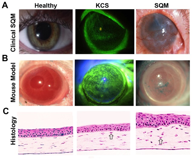
Figure 1. Keratoconjunctivitis sicca (KCS) and squamous metaplasia (SQM) of the ocular surface in response to CD4+ T cell-mediated autoimmunity.
Representative images of autoimmune-mediated ocular surface disease in a (A) human patient and (B) Aire KO mouse. Aqueous tear deficiency leads to KCS with loss of epithelial integrity indicated by punctate fluorescein staining (A&B-middle panels). SQM is accompanied by pathological keratinization, corneal opacification and vascularization (A&B - right panels). (C) H&E staining of cryosectioned eyes from Aire KO mice reveals representative histological changes associated with disease progression. Open arrows indicate infiltrating immune cells in the corneal stroma.

