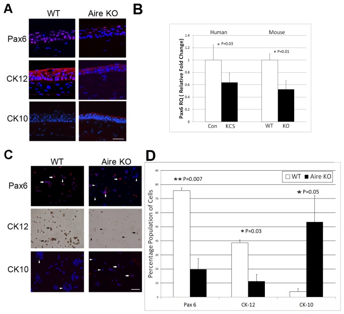
Figure 2. Altered lineage commitment of the ocular mucosal epithelium in autoimmune-mediated KCS/SQM.
(A) Immunofluorescent staining of nuclear Pax6 (red, top), corneal-specific CK12 (red, middle) and epidermal-specific CK10 (red, bottom) in the corneal epithelium of WT and Aire KO mice. Scale bar = 50μm (B) TaqMan qPCR analysis of Pax6 gene using corneolimbal epithelial cells from WT and Aire KO mice and impression cytology specimens from healthy human control (Con) and chronic KCS (KCS) patients. Pax6 expression in WT mice or healthy control patients was used as the reference (designated 1-fold) to generate relative quantitation (RQ) values. Data are shown as mean RQ±SE. Seven mice were studied per group, as well 15 KCS patients and 7 healthy controls. Unpaired T-test (mouse) or Wilcoxan Rank Sum test (human) was used to test for differences between groups, with P<0.05 (*) and P<0.01 (**) considered statistically significant. (C) Cytospin was used to assess single-cell expression of Pax6 (red, upper panels), CK12 (brown, middle panels) and epidermal cytokeratin K10 (red, bottom panels) in WT and KO mice. Arrows and arrowheads indicate positive and negative stained cells, respectively. (D) Quantitative analysis of cytospin specimens expressed as the percentage of cells staining positive for Pax6, CK12 and CK10. Data were generated by analyzing three separate fields of view from an individual specimen with a minimum of three animals analyzed per group. Group mean comparisons were assessed by T-test, with P<0.05 (*) and P<0.01 (**) considered statistically significant.

