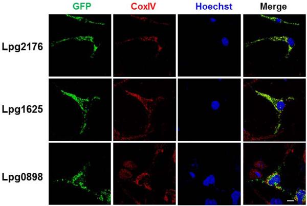
Fig. 2. Some of the caspase-3 activators localized to the mitochondrion.
Hela cells transfected to express GFP fusion of the indicated proteins were stained with antibody specific for the mitochondrial protein COX4I1. Samples were analyzed using an Olympus IX-81 fluorescence microscope for images acquisition. Images were pseudocolored with the IPLab software package. Bar: 20 μm.

