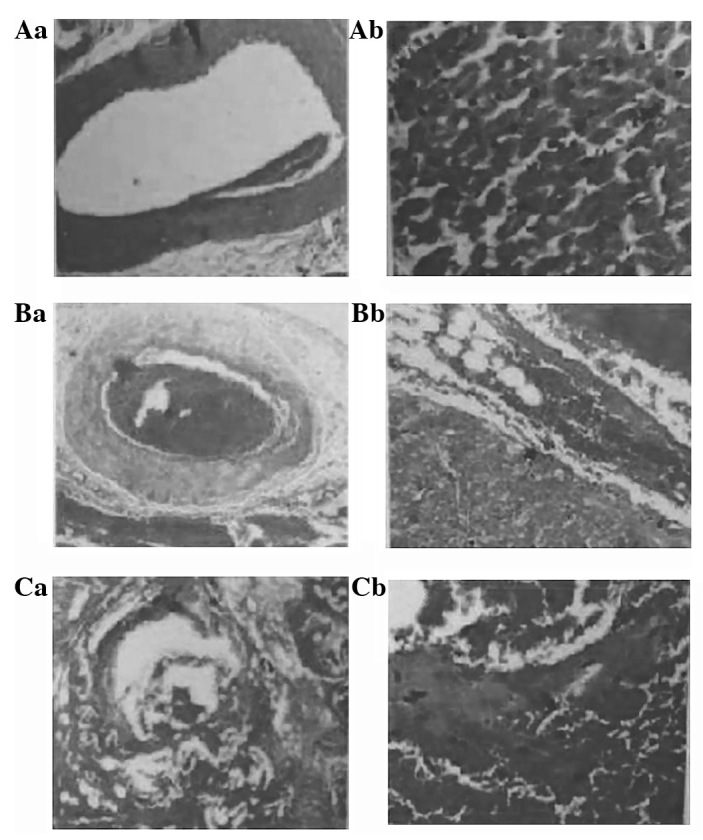
Figure 5.

Pathological findings following percutaneous transluminal radio-frequency closure (PTRFC). Protected sections; (Aa) the blood vessel and (Ab) the myocardium are normal. Thrombus section: (Ba) mixed thrombus engorgement, blocked vessel cavities in the epicardium and myocardial layer, destroyed inner membranes of blood vessels, and atrophic smooth muscles of the middle membrane; (Bb) incomplete blockage of a few small blood vessels, swollen neighboring myocardial cells, and granular degeneration in the cell plasma. A section of the myocardium was swollen, had merged together, and resembled ‘hot, solidifying’ necrosis. Damaged section: (Ca) almost complete destruction of the vessel wall structure. (Cb) Bleeding between the neighboring myocardium, swollen myocardial cells with disappearing horizontal lines, dissolved and disappearing nuclear breaks, and myocardial necrosis.
