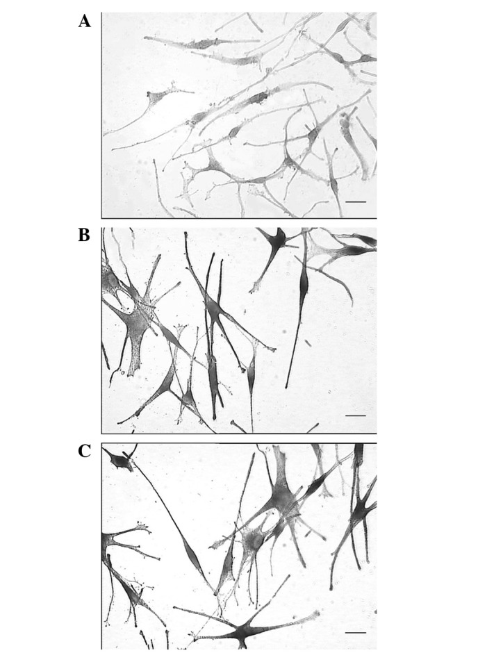
Figure 4.

(A) No black granules are visible in the cytoplasm of the melanocyte precursors (MPs). The cytoplasm is stained by the nuclear fast red counterstain. (B) Following treatment with 1,25-dihydroxyvitamin D3 (VID), black granules are visible in the MPs, and the majority of the cells show strong 3,4-dihydroxy-L-phenylalanine (DOPA) staining, which obscures the nuclear fast red staining. (C) The results in the melanocytes are similar to those in the VID-treated MPs, showing strong positive staining and black granules that obscure the nuclear fast red staining. (DOPA stain, nuclear fast red counterstain; scale bar, 50 μM.)
