Figure 2.

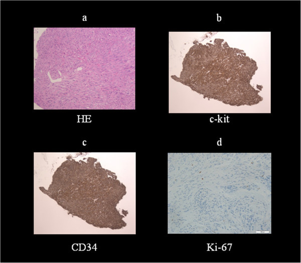
Histopathological findings. a) Spindle cell tumor b,c) Positive immunostaining for c-kit and CD34 d) <3% staining for Ki-67 antigen, a cellular proliferation marker.

Histopathological findings. a) Spindle cell tumor b,c) Positive immunostaining for c-kit and CD34 d) <3% staining for Ki-67 antigen, a cellular proliferation marker.