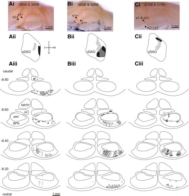
Figure 6.
Retrograde cell labeling in the pons. Ai, Photomicrograph of posterior lobe of the rat cerebellum showing red and green Retrobeads injections in e1+ and e2+, respectively. Aii, Standard horizontal map of vDAO showing territory occupied by olive cells retrogradely labeled with green Retrobeads (light gray) and red Retrobeads (black). Aiii, Distribution of retrogradely labeled pontine cells plotted on standard transverse maps (0.2 mm interval between maps, AP levels −8.2 to −8.8). Each symbol indicates a labeled cell (black crosses denote red-labeled cells, gray circles denote green-labeled cells). Bi–Biii, Same as Ai–Aiii but the red and green injection sites were both located in zebrin band e1+. Ci–Ciii, Same as Ai–Aiii but the red injection site was located in zebrin bands e1− and e2+ and the green injection site was located in zebrin band e2+ with spread to neighboring e2−. BPN, Basilar pontine nuclei; c, caudal; l, lateral; m, medial; ml, medial lemniscus; ped, cerebral peduncle; NRTP, nucleus reticularis tegmenti pontis; r, rostral.

