
Figure 8.
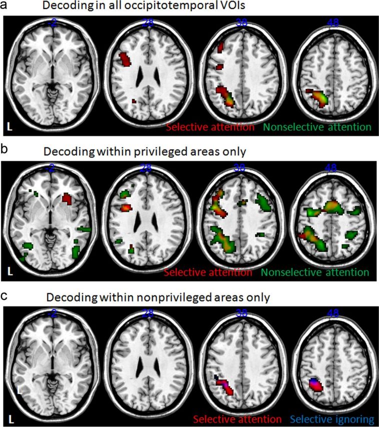
Correlations between occipitotemporal decoding ability and whole-brain fMRI activity. Areas showing a significant effect (uncorrected voxel-level p < 0.001, FWE-corrected cluster level p < 0.05) in a voxel-based one-sample t test across all subjects. Input images were contrast values reflecting the partial correlation between trial-by-trial decoding probability for selectively ignored (blue), selectively attended (red), and nonselectively attended (green) trials and the fMRI signal in that voxel. All effects shown are positive (correlations >0); there were no significant negative correlations. The different rows reflect different occipitotemporal areas that were used to calculate decoding probabilities: a, decoding based on all occipitotemporal areas; b, decoding performed within the occipitotemporal area thought to be privileged for the category of interest (privileged area regressors; see Materials and Methods); c, decoding within the nonprivileged regions (nonprivileged area regressors; see Materials and Methods).