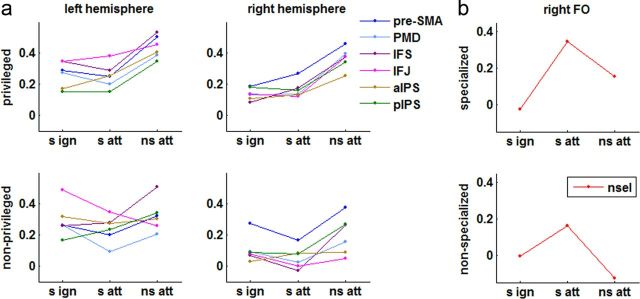
Figure 9.
Correlations between occipitotemporal decoding ability and frontal and parietal fMRI activity. Plotted are the contrast values reflecting the partial correlation between trial-by-trial decoding probability for selectively ignored (s ign), selectively attended (s att), and nonselectively attended (ns att) trials and the fMRI signal in a given VOI (for exact VOI coordinates, see Table 1). The top row shows the results when decoding was performed within the occipitotemporal area thought to be privileged for the category of interest (privileged area regressors; see Materials and Methods); the bottom row shows results when decoding within the remaining nonprivileged regions (nonprivileged area regressors; see Materials and Methods). a, Frontal and parietal areas defined based on the nonselective attend contrast (Fig. 8b, green). Areas are color coded and plotted separately for the left and right hemispheres. b, Right FO VOI as defined based on the selective attend contrast (Fig. 8b, red).

