
Figure 4.
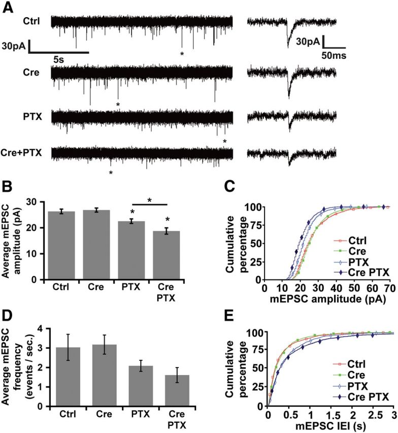
NF-κB opposes the homeostatic downregulation of synaptic strength by elevated activity. A, Representative traces of mEPSCs recorded from p65-wild-type or p65-deficient (Cre) DIV21 RelAF/F hippocampal cultures after treatment with vehicle or PTX (100 μm for 20–28 h). Right, Enlarged single events (marked by asterisks) from each condition. B, NF-κB limits the homeostatic decrease in AMPAR-dependent mEPSC amplitudes in response to elevated activity in hippocampal neurons. Average AMPAR-dependent mEPSC amplitudes under the indicated conditions (*p ≤ 0.023, ANOVA). C, Cumulative percentage plot of mEPSC amplitudes from all recorded neurons (nCtrl = 12, nCre = 10, nPTX = 10, nCre+PTX = 11, where n represents individual neurons recorded from 10 independent experiments). K–S statistical analysis shows a significant left shift in the cumulative distribution of mEPSC amplitudes from Ctrl after chronic PTX both in the presence (PTX; p = 0.014) and absence of p65 (Cre/PTX; p = 9.67 × 10⁻⁴). There is also a statistically significant left shift in the cumulative distribution of mEPSC amplitudes in neurons treated with PTX in the absence of p65 (Cre/PTX) compared to neurons with p65 (PTX; p = 8.65 × 10⁻³). D, Average AMPAR-dependent mEPSC frequency under the indicated conditions. E, Cumulative percentage plot of mEPSC IEIs from all recorded neurons. K–S test shows statistically significant right shifts from Ctrl in the cumulative distribution of mEPSC IEIs after chronic PTX in the absence of p65 (Cre/PTX; p = 0.018), and between chronic PTX in the presence (PTX) and absence of p65 (Cre/PTX; p = 0.035), but there is no statistical difference from Ctrl in the presence of PTX (PTX; K–S test). All error bars indicate SEM.